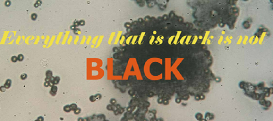
An appeal from Mucormycosis: Do not call me Black Fungus
The pandemic has educated us with a bunch of new terminologies with the addition of black fungus to the new terror list. First of all, these demons have always resided in microbiology textbooks and acted like a terror for Microbiology students like us. So, they are nothing new. There are Zillions of viruses, bacteria, fungi, and parasites around to nail us to death. They were always there in textbooks, research papers, and clinical notes, it’s just that the pandemic has brought them to newspapers and TV channels. Thanks to mother nature and our immune system we are still alive even with a co-existence with them. Once the immune mechanism is compromised we become prey to those microscopic demons.
*We are not the same*
However,
the preface is just to clear confusion about the overlap of two terminologies
in news these days. The terms Black fungus disease and mucormycosis are being
used interchangeably in popular articles and media. *Unfortunately, these two
are entirely different disease that shares some similarities in the
pathophysiology and presentation.* The prominent point that misguided us is their
ability to attack immunocompromised patients at more or less similar target
organs as in the case of COVID-19 infection. Moreover, no scientific literature
has mentioned the term “ Black fungus” while describing research on
mucormycosis and vice versa. Let us have a quick look at some dissimilarities of
these two diseases:
*Different etiological agents*
Blank
fungi or sometimes known as black yeasts fall into the class of Dematiaceous
fungi that are responsible for a wide variety of infectious syndromes. They are
often found in soil and rotten plant materials and are generally inhaled by most if
not all individuals. However, its pathogenicity becomes significant in
immunocompromised individuals.
The
clinical nomenclatures caused by these black fungi are mycetoma,
chromoblastomycosis, phaeohyphomycosis, and NOT mucormycosis which is of
entirely different origin. The causative genera for these clinical
manifestations are Exophiala dermatitidis, Cladophialophora-, Fonsecaea- and
Ramichloridium-like strains, known in humans as agents of chromoblastomycosis.
On
the other hand, the causative agents for mucormycosis are certain fungi,
especially Rhizopus and mucormycetes, which are common contaminants of
laboratory cultures. Some of the fungi that are responsible for mucormycosis
are Rhizopus species, Mucor species, Rhizomucor species, Syncephalastrum
species, Cunninghamella bertholletiae, Apophysomyces species, and Lichtheimia
(formerly Absidia) species.
*What is BLACK in it*
At
this point, this is clear that the causative agents are completely different
for these two types of diseases. Let us
take a break from finding the differences between these two clinical
manifestations and concentrate on a more interesting question.
*Why
these Dematiaceous fungi are called “BLACK” fungus?*
Interestingly,
these fungi possess unique pathogenic mechanisms attributed to the presence of
melanin in their cell walls, which imparts the characteristic dark color to
their spores and hyphae. On contrary, no such characteristic features are
consistently prominent in the microscopic observation of all fungal species that
are mucormycosis-causing organisms. Of note, Mucormycosis causes tissue
blackening by devitalizing its blood supply, and this black appearance has been
misinterpreted, misreported, and misdiagnosed as a black fungus by the media.
*We are on the right path*
Fortunately,
despite the confusing terminology, our able physicians have detected the
correct disease in COVID-affected individuals- it is Mucormycosis NOT anything
caused by black fungus. Therefore, they are predominantly treated by IV amphotericin
B and some azoles. Of note, itraconazole and voriconazole demonstrate the most
consistent in vitro activity against black fungi (except S. prolificans, which
are resistant to azoles) whereas voriconazole is completely ineffective in
mucormycosis. Whereas, fluconazole and echinocandins exhibit limited or no
activity against both types of diseases.
There
are many other similarities in the pathogenesis and presentation that lead to
the overlap of the terminologies. Sources for both the etiological agents are
the same: soil, composts, rotten woods, and decaying organic matter. Mucormycosis
has rhinocerebral, ocular, pulmonary, and gastrointestinal involvements. The blank
fungus affects similar organs with less severity than mucormycosis.
Mucormycosis is a more morbid condition than diseases involving black
fungus. It often requires multiple surgeries even involving enucleation (Complete
removal of the eye).
*Final Words*
I
am not discussing the co-morbidities that trigger the mucormycosis infection as
it is now a well-known topic. Just to mention that there is no scientific
evidence that oxygen pipes, oxygen flowmeter, or humidifier are responsible for
its spread or origin. Unlike Covid-19, it is not contagious and does not spread
from one person to another. Finally, the causative agents are co-existing with
us in nature and it is not going to be fatal to you unless you are being
treated with high steroids and your diabetes is not under control during your
COVID infections days.
References
Baker, R. D. (1957).
Mucormycosis—a new disease?. Journal of the American Medical
Association, 163(10), 805-808.
Baker
R.D. (1971) Mucormycosis. In: The Pathologic Anatomy of Mycoses. Handbuch der
Speziellen Pathologischen Anatomie und Histologie (Atmungswege und Lungen), vol
3 / 5. Springer, Berlin, Heidelberg. https://doi.org/10.1007/978-3-642-80570-7_22
Hoog,
G. S. de, Queiroz-Telles, F., Haase, G., Fernandez-Zeppenfeldt, G., Angelis, D.
A., H. G. Gerrits van den Ende, A., … Yegres, F. (2000). Black fungi: clinical
and pathogenic approaches. Medical Mycology, 38(s1), 243–250.
doi:10.1080/mmy.38.s1.243.250
McGinnis MR, Hilger AE.
Infections Caused by Black Fungi. Arch Dermatol. 1987;123(10):1300–1302.
doi:10.1001/archderm.1987.01660340062020
https://www.cdc.gov/fungal/diseases/mucormycosis/treatment.html
Revankar SG.
Dematiaceous fungi. Mycoses. 2007 Mar;50(2):91-101. doi:
10.1111/j.1439-0507.2006.01331.x. PMID: 17305771.


